
DOI: 10.5281/zenodo.20473791 · UDC: 618.36-089.85
Introduction. Ovarian ectopic pregnancy is a rare form of ectopic implantation associated with a high risk of rupture and life-threatening intra-abdominal hemorrhage. Preoperative diagnosis remains difficult because of the nonspecific clinical and ultrasonographic findings, especially in cases with atypical presentation.
Case presentation. We present the case of a 26-year-old woman with a 3-year history of infertility who presented with epigastric pain and diarrhea in early pregnancy. Transvaginal ultrasonography revealed the absence of an intrauterine gestational sac, a left adnexal mass, and significant hemoperitoneum. Emergency laparoscopy identified a ruptured left ovarian ectopic pregnancy complicated by massive hemoperitoneum of approximately 2400 mL. Partial ovarian resection with removal of trophoblastic tissue and preservation of viable ovarian parenchyma was performed successfully.
Conclusions. This case highlights the importance of maintaining a high index of suspicion for ectopic pregnancy in women of reproductive age presenting with atypical abdominal or gastrointestinal symptoms. Early diagnosis and prompt minimally invasive surgical management are essential for preventing severe maternal morbidity and preserving reproductive potential.
Ectopic pregnancy is one of the most serious gynecological emergencies, due to the risk of internal bleeding, hemodynamic compromise, adverse effects on reproductive prognosis, and, in the absence of prompt diagnosis and treatment, even maternal death [1, 2]. In current practice, the diagnosis of ectopic pregnancy is based primarily on the correlation between transvaginal ultrasound and dynamic monitoring of β-hCG (beta-human chorionic gonadotropin), but diagnostic difficulties persist, especially in non-tubal forms and in cases with atypical clinical presentation [1, 3]. In this context, ovarian pregnancy occupies a special place, being one of the rarest locations of ectopic pregnancy, but also one of the most difficult to recognize preoperatively [4-6].
The relevance of this topic stems primarily from the rarity of ovarian pregnancy and its severe clinical implications. According to recent data, ovarian pregnancy accounts for approximately 0.5-3% of all ectopic pregnancies, which explains the limited experience many clinicians have in recognizing and managing this condition [4-8]. Although rare, this condition is disproportionately significant, as its course can rapidly be complicated by ovarian rupture and hemoperitoneum, necessitating emergency surgery [4, 6, 8]. Furthermore, the current literature indicates that preoperative diagnosis remains difficult, and definitive confirmation is frequently established intraoperatively and histopathologically [4, 5, 7]. This case gains additional clinical significance from the patient’s history of infertility, as recent literature highlights the role of the endometrial microbiome in maintaining uterine homeostasis, endometrial receptivity, and the inflammatory and immunological mechanisms involved in infertility, thereby supporting a comprehensive reproductive assessment in patients with rare ectopic pregnancies, including ovarian pregnancy [9].
The difficulty of differential diagnosis is another key reason why this topic is important. Clinically, an interrupted ovarian pregnancy may present with acute pelvic pain, short-term amenorrhea, and minimal or absent genital bleeding — a presentation that can mimic a ruptured tubal pregnancy, a hemorrhagic corpus luteum cyst, ovarian apoplexy, adnexal torsion, or even an incomplete spontaneous abortion [1, 4, 5]. Consequently, clinical suspicion is often nonspecific, and accurately determining the site of implantation becomes a real diagnostic challenge [4-6]. It is precisely this clinical and imaging overlap with other gynecological emergencies that justifies the increased scientific interest in the analysis of cases of interrupted ovarian pregnancy.
The relevance of this subject is further increased by the fact that some forms of ectopic pregnancy may present atypically, with gastrointestinal symptoms or signs mimicking acute intestinal pathology. The literature describes cases of abdominal, tubal, or retroperitoneal ectopic pregnancy in which the initial diagnosis was missed or misdirected toward acute digestive disorders, including gastroenteritis, against a background of severe abdominal pain, epigastric localization of pain, diarrhea, syncope, or other nonspecific manifestations [10-12]. These clinical presentations may delay the recognition of an obstetric hemorrhagic emergency, especially when classic gynecological signs are absent or mild [13]. Thus, the presence of signs suggestive of intestinal dysfunction, such as epigastric pain, diarrhea occurring shortly before seeking medical care, and dehydration syndrome, does not rule out a complicated ectopic pregnancy, but rather necessitates maintaining a high clinical suspicion in any woman of reproductive age with amenorrhea and unexplained abdominal or gastrointestinal symptoms [10-12].
Imaging diagnosis of ovarian pregnancy has made significant progress in recent years, but remains imperfect. Transvaginal ultrasound is the first-line method for diagnosing ectopic pregnancy [1, 3, 5], and in the case of ovarian pregnancy, suggestive signs may include the presence of a gestational sac located in the ovarian cortex, sometimes with a yolk sac or embryo, as well as the inability to separate the mass from the ovary upon gentle palpation with the vaginal probe [5, 6]. However, these signs are not always evident, and the lesion can easily be confused with a hemorrhagic corpus luteum or a complex adnexal mass [5, 6]. Recent data indicate that, in centers with expertise in early pregnancy ultrasound, most cases can be identified preoperatively, allowing for better treatment planning and a minimally invasive approach [6].
This topic remains relevant because an interrupted ovarian pregnancy continues to pose challenges regarding the confirmation of a definitive diagnosis. Despite advances in imaging, Spiegelberg’s classic criteria remain relevant for distinguishing primary ovarian pregnancy from other forms of ectopic implantation with secondary ovarian involvement [4, 7]. These criteria include the integrity of the fallopian tube on the same side, the location of the gestational sac in the ovarian region, the connection of the ovary to the uterus via the ovarian ligament, and histological evidence of ovarian tissue in the wall of the gestational sac [7]. The continued reliance on intraoperative and histopathological confirmation demonstrates that missed ovarian pregnancy remains a current diagnostic challenge, insufficiently standardized in current clinical algorithms [4, 5, 7].
Therapeutic implications further emphasize the importance of this topic. In modern gynecology, the management of ectopic pregnancy aims not only to save the patient’s life but also to preserve reproductive function whenever possible [1, 2]. The treatment of ovarian pregnancy is, in most cases, conservative surgical management, preferably by laparoscopy, involving excision of trophoblastic tissue or partial ovarian resection with hemostasis and maximal preservation of healthy ovarian tissue; methotrexate may be considered only in early, unruptured cases, in hemodynamically stable patients with the possibility of strict follow-up [14]. Treatment for interrupted ovarian pregnancy is most often surgical, and laparoscopy is preferred when the patient’s clinical condition permits it [4, 7]. Conservative intervention, involving excision of the trophoblastic tissue and maximal preservation of healthy ovarian parenchyma, is of particular importance in young women and in patients with unfulfilled reproductive potential [4, 6, 7]. In contrast, in cases complicated by significant bleeding or hemodynamic instability, a more extensive procedure may be necessary [1, 4]. Thus, the clinical assessment of an interrupted ovarian pregnancy has immediate practical relevance for choosing the optimal management strategy and for balancing radicality with the preservation of fertility.
In addition, the issue is supported by the fact that there's growing interest in identifying risk factors and understanding the pathophysiological characteristics of ovarian implantation. Recent data suggest that the risk factors for ovarian pregnancy partially overlap with those for ectopic pregnancy in general; however, the use of intrauterine devices (IUDs) and assisted reproductive techniques (ART) appears to be reported more frequently in this rare form of implantation [4, 8]. At the same time, some recent clinical analyses have identified associations with ART and IUDs as potential contributing factors, highlighting that patients with ovarian pregnancy may develop more severe clinical complications [8]. Given the increasing use of modern reproductive technologies and the intensification of early pregnancy monitoring, the recognition of rare ectopic locations is becoming increasingly important for current obstetric and gynecological practice.
From a scientific viewpoint, the topic remains relevant due to the lack of large, prospective studies; most of the evidence consists of case reports, small series, and systematic reviews based on heterogeneous literature [4, 7]. A systematic review published in 2023 emphasizes that, although ovarian pregnancy is a well-known condition, its pathophysiological mechanisms, optimal diagnostic criteria, and therapeutic options are not yet fully standardized [7]. For this reason, every rigorously documented case of interrupted ovarian pregnancy has real scientific value, contributing to the refinement of diagnostic criteria, the optimization of the surgical approach, and the consolidation of collective clinical experience in a rare but potentially severe condition [4, 6, 7].
Finally, the relevance of this topic stems from the need to reduce preventable maternal morbidity. Ectopic pregnancy remains a major cause of severe complications in the first trimester, and rare forms, such as ruptured ovarian pregnancy, are particularly dangerous when diagnosis is delayed [1, 3, 4]. The rich vascularization of the ovary promotes rapid intra-abdominal hemorrhage in the event of rupture, which can turn this rare condition into a life-threatening emergency [4]. Ovarian pregnancy, as a rare form of ectopic pregnancy, requires early diagnosis and individualized treatment to prevent maternal hemorrhagic complications, while subsequent reproductive management requires careful assessment of the obstetric risks associated with future pregnancies [15]. In this context, the development and publication of studies dedicated to ovarian pregnancy are fully justified, as they contribute to increased clinical vigilance, improved ultrasound recognition, and the timely selection of an appropriate therapeutic approach [4-6].
Despite advances in diagnosis and the management of ectopic pregnancy, this condition remains insufficiently standardized, and the current literature supports the need for systematic reporting and analysis of clinical cases [4, 6, 7]. The study of this condition thus has clear practical and scientific relevance for obstetricians-gynecologists, radiologists, emergency physicians, and pathologists involved in the diagnosis and treatment of emergencies in early pregnancy, which is why we present this case.
On March 13, 2026, at 11:30 p.m., a 26-year-old patient, G1P0, with a 3-year history of infertility, presented to the emergency department of the “Gheorghe Paladi” Municipal Clinical Hospital in Chișinău, Moldova. She had initially sought medical assistance via the 112-emergency service for diarrhea and epigastric pain. She was initially examined by the infectious disease specialist at the infectious disease’s hospital in Chișinău. Given a known pregnancy of approximately 5 weeks, the infectious disease specialist recommended a gynecological evaluation to rule out an acute gynecological cause. According to medical history, pregnancy occurred spontaneously after approximately 3 years of infertility. No drug allergies or previous gynecological procedures were noted in the records provided.
The patient was afebrile and in moderately serious condition upon admission to the emergency department. Her hemodynamic parameters were borderline (blood pressure approximately 100/65 mmHg). The abdomen was moderately tender on palpation. The clinical presentation raised suspicion of acute gynecological abdomen with possible intraperitoneal hemorrhage.
Laboratory tests revealed a serum β-hCG level of 9,544.7 mIU/mL, which was measured on the patient's own initiative, in the morning on March 13, 2026. The complete blood count revealed signs of anemia, with hemoglobin at 86 g/L, red blood cells at approximately 2.8×10¹²/L, and a hematocrit of 0.26. The platelet count was approximately 130×10⁹/L, and the white blood cell count was approximately 8.1×10⁹/L. Assessment of hemostasis during the peri- and postoperative periods showed a prothrombin activity (Quick) of 65.1%, an INR of 1.33, and a fibrinogen level of 2.53 g/L.
Transvaginal ultrasound did not reveal an intrauterine pregnancy. A mass suggestive of an ectopic pregnancy was identified in the left adnexa, associated with the presence of free intraperitoneal fluid and clots – an ultrasound finding suggestive of significant hemoperitoneum. Based on the clinical and laboratory findings, a presumptive diagnosis of ectopic pregnancy complicated by hemoperitoneum was made. Given the suspicion of intraperitoneal hemorrhage and the risk of decompensation, emergency surgery was indicated.
On 14 March 2026, at 00:05, the patient underwent emergency laparoscopic surgery, comprising partial resection of the left ovary with removal of the gestational tissue, achievement of hemostasis, and drainage of the peritoneal cavity (figure 1).
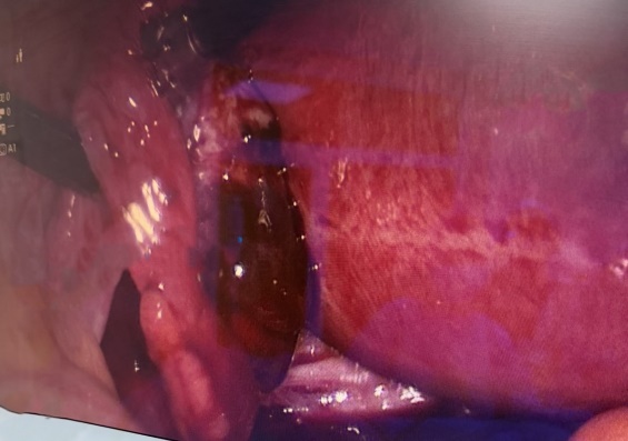
The procedure was performed under general anesthesia, following establishment of a CO₂ pneumoperitoneum. Laparoscopic exploration of the abdominal cavity revealed a massive hemoperitoneum, with the presence of both liquid blood and clots; approximately 2,400 mL was aspirated. The uterus was of normal size and configuration. Both Fallopian tubes appeared macroscopically normal, with preserved anatomical integrity and free fimbriae. The right ovary was unremarkable. The left ovary was found to be adherent to the posterior leaf of the broad ligament and embedded within blood clots; on further inspection, a purplish mass measuring approximately 2 × 3 cm was identified, with an actively bleeding perforation site suggesting the site of ectopic implantation.
In view of the intraoperative findings, partial resection of the left ovary was undertaken, with local excision of the pathological tissue and removal of the products of conception, while preserving the remaining viable ovarian parenchyma. Hemostasis was secured by diathermocoagulation, achieving complete control of active bleeding. At the conclusion of the procedure, two transcutaneous drains were placed in the pouch of Douglas. The surgical specimen, consisting of ovarian tissue associated with products of conception, was submitted for histopathological examination.
Given the massive blood loss and severe post-hemorrhagic anemia, perioperative management included volume resuscitation and transfusion support, with packed red blood cells administered both intraoperatively and postoperatively. Determination of the patient’s blood group, A(II) Rh positive, was also incorporated into perioperative care. Hemostatic assessment showed moderately reduced prothrombin activity (Quick 65.1%) and an international normalized ratio (INR) of 1.33, with a fibrinogen concentration of 2.53 g/L and no evidence of hypofibrinogenemia. Overall, these findings were compatible with mild coagulation disturbance in the context of acute hemorrhage and hemodilution.
The postoperative course was favorable, with close clinical and para-clinical surveillance. The postoperative diagnosis was interrupted/arrested left ovarian ectopic pregnancy complicated by hemoperitoneum and severe post-hemorrhagic anemia, in the setting of intraoperative and postoperative packed red blood cell transfusion. Postoperative drainage yielded a small volume of serosanguineous fluid. Serial full blood count assessment demonstrated progressive improvement in hematological parameters, with hemoglobin reaching approximately 99 g/L, hematocrit – 0.29, and an erythrocyte count of 3.2×10¹²/L. A moderate leukocytosis (approximately 13.5 × 10⁹/L), accompanied by neutrophilia and a left shift of the leukocyte formula (band neutrophils approximately 13% and segmented neutrophils approximately 71%), was also observed, consistent with a postoperative inflammatory response. β-hCG measured on 16 March 2026 was 1163.67 mIU/mL, representing a significant decline from the initial value and supporting effective removal of trophoblastic tissue. The patient was discharged on postoperative day 3, with recommendations for antibacterial, anti-inflammatory, and analgesic therapy, together with ongoing clinical and laboratory follow-up by her general practitioner. At discharge, the patient provided written informed consent for publication of this case.
As noted in the literature, the preoperative recognition of ovarian ectopic pregnancy remains challenging despite recent advances in early pregnancy ultrasonography [4, 6-8]. The present case confirms these observations, highlighting the misleading nature of the initial clinical presentation, which was dominated by gastrointestinal manifestations, namely diarrhea and epigastric pain. The literature indicates that certain forms of ectopic pregnancy may present atypically, mimicking acute gastrointestinal pathology, thereby initially directing assessment towards other specialties and delaying recognition of an obstetric hemorrhagic emergency [10-12]. In this context, the case underscores the importance of maintaining clinical suspicion for ectopic pregnancy in any woman of reproductive age with a known pregnancy or amenorrhea who presents with abdominal pain, even when gynecological symptoms are subtle or absent [1, 3].
As noted, preoperative diagnosis is often hindered by clinical and imaging similarities to other adnexal emergencies [4-7]. In the present case, however, the absence of an intrauterine gestational sac on transvaginal ultrasonography, together with the presence of free intraperitoneal fluid and blood clots, raised a strong suspicion of ectopic pregnancy complicated by hemoperitoneum. Confirmation of the ovarian location was achieved intraoperatively, through identification of bilaterally normal fallopian tubes and a hemorrhagic lesion localized to the left ovary, a finding consistent with the classical criteria used for the diagnosis of primary ovarian pregnancy [4, 7]. This is in keeping with recent literature, which shows that, in many cases, definitive confirmation remains intraoperative and histopathological [4, 5, 7].
A particularly relevant feature of this case is the severity of the intra-abdominal hemorrhage. The hemoperitoneum volume, approximately 2,400 mL, accounts for the severe post-hemorrhagic anemia and the perioperative transfusion requirement. The literature emphasizes that, although rare, ovarian pregnancy may rapidly become complicated by rupture and massive hemorrhage because of the ovary’s rich vascular supply, thereby turning the clinical picture into a life-threatening emergency [4, 6, 8]. From this perspective, the case illustrates an important practical lesson: apparently relatively preserved hemodynamic stability at presentation does not exclude severe intraperitoneal hemorrhage, and the association of early pregnancy, abdominal pain, and free intraperitoneal fluid should be interpreted with the utmost vigilance [1, 3, 4].
The surgical management applied in this case reflects current principles in the treatment of complicated ovarian ectopic pregnancy. Whenever the patient’s clinical condition permits, laparoscopy is considered the preferred approach, as it provides both accurate diagnosis, evacuation of the hemoperitoneum, hemorrhage control, and the opportunity to preserve reproductive function [4, 6, 7]. In the present case, the laparoscopic intervention allowed both confirmation of the diagnosis and performance of a partial ovarian resection with excision of the trophoblastic tissue while preserving the remaining viable ovarian parenchyma. This represents one of the main strengths of the management strategy, given the patient’s young age, her 3-year history of infertility, and the fact that the pregnancy had been achieved spontaneously. In the literature, maximal preservation of healthy ovarian tissue is regarded as particularly important in patients with unfulfilled reproductive potential [4, 6, 7].
The postoperative course and the dynamics of the biological markers supported the effectiveness of the adopted management. The significant postoperative decline in β-hCG was consistent with effective removal of the trophoblastic tissue, while the gradual improvement in hematological parameters confirmed a favorable response to both surgical and supportive treatment. In addition, monitoring coagulation parameters had important practical relevance. In the setting of massive hemorrhage, assessment of Quick value, INR, and fibrinogen allows early evaluation of the impact of acute blood loss and hemodilution, as well as guidance of transfusion support [1, 3]. In the present case, the moderate reduction in prothrombin activity and slight increase in INR, with fibrinogen remaining within acceptable limits, suggested early coagulation impairment without the development of severe coagulopathy, thus allowing therapeutic control to be maintained.
This case is instructive for several reasons. Firstly, it demonstrates that gastrointestinal symptoms do not exclude a severe gynecological cause in early pregnancy [9-11]. Secondly, it highlights the decisive utility of transvaginal ultrasonography correlated with β-hCG in the rapid diagnostic orientation of ectopic pregnancy [1, 3, 5, 6]. Thirdly, it shows that laparoscopy may represent not only an effective method of controlling life-threatening hemorrhage, but also a fertility-preserving option in carefully selected cases [4, 6, 7]. Therefore, the principal lesson drawn from this case is the need for heightened clinical vigilance and rapid multidisciplinary reassessment in pregnant women in the first trimester presenting with atypical abdominal pain, to avoid delayed diagnosis and worsening hemorrhagic complications [1, 3, 4].
This case illustrates that ovarian ectopic pregnancy, although rare, may lead to massive hemoperitoneum and severe post-hemorrhagic anemia, with rapid progression to a life-threatening condition. The atypical presentation, dominated by gastrointestinal symptoms, underscores the need to prioritize exclusion of ectopic pregnancy in any first-trimester patient presenting with abdominal pain. In the presence of signs of intraperitoneal bleeding, prompt surgical intervention is essential. In this case, laparoscopy enabled both rapid hemorrhage control and ovarian-conserving treatment, an aspect of particular importance given the patient’s young age, history of infertility, and unfulfilled reproductive potential.
Hendriks E, Rosenberg R, Prine L. Ectopic Pregnancy: Diagnosis and Management. Am Fam Physician. 2020;101(10):599-606.
American College of Obstetricians and Gynecologists' Committee on Practice Bulletins—Gynecology. ACOG Practice Bulletin No. 193: Tubal Ectopic Pregnancy. Obstet Gynecol. 2018;131(3): e91-e103.
Mullany K, Minneci M, Monjazeb R, C Coiado O. Overview of ectopic pregnancy diagnosis, management, and innovation. Womens Health (Lond). 2023;19:17455057231160349. doi:10.1177/17455057231160349
Bouab M, Touimi AB, Jalal M, Lamrissi A, Fichtali K, Bouhya S. Diagnosis and management of ectopic ovarian pregnancy: a rare case report. Int J Surg Case Rep. 2022;91:106742. doi:10.1016/j.ijscr.2021.106742.
International Society of Ultrasound in Obstetrics and Gynecology. Ovarian ectopic pregnancy. ISUOG. Published 2022. Accessed March 18, 2026. https://www.isuog.org/clinical-resources/patient-information-series/patient-information-pregnancy-conditions/early-pregnancy/ovarian-ectopic-pregnancy.html
Solangon SA, Naftalin J, Jurkovic D. Ovarian ectopic pregnancy: clinical characteristics, ultrasound diagnosis and management. Ultrasound Obstet Gynecol. 2024;63(6):815-823. doi:10.1002/uog.27549
Almahloul Z, Amro B, Nagshabandi Z, et al. Ovarian Pregnancy: 2 Case Reports and a Systematic Review. J Clin Med. 2023;12(3):1138. doi:10.3390/jcm12031138
Li H, Liu Y, Yang Y, Zhao X, Qi X. Clinical analysis of women with ovarian pregnancy: a retrospective case-control study. BMC Pregnancy Childbirth. 2022;22(1):768. Published 2022 Oct 13. doi:10.1186/s12884-022-05099-8
Burac M, Surguci M, Corolcova N, Cotelea V, Mihalcean L, Caproș H. Microbiomul endometrial în infertilitate: roluri, mecanisme şi implicaţii clinice = The endometrial microbiome in infertility: roles, mechanisms, and clinical implications. Moldovan Journal of Health Sciences. 2025;12(3 Suppl 2):518.
Brandt AL, Tolson D. Missed abdominal ectopic pregnancy. J Emerg Med. 2006;30(2):171-174. doi:10.1016/j.jemermed.2005.04.018
Restaino S, Degano M, Padovani D, et al. A Case of Advanced Tubal Ectopic Pregnancy after Emergency Contraception. Healthcare (Basel). 2022;10(8):1590. doi:10.3390/healthcare10081590
Xia L, Qi T, Qian J. A Case Report of Retroperitoneal Ectopic Pregnancy and Review of Literature. Int J Womens Health. 2024;16:1855-1864. doi:10.2147/IJWH.S486185
Mihalcean L, Corolcova N, Burac M, Cotelea V. Ectopic pregnancy associated with the use of a levonorgestrel-releasing intrauterine system (LNG-IUS, JAYDESS): case report and clinical implications. MEDICUS. 2026;(1):13-16.
Surguci M, Ivas T. Utilizarea metotrexatului în sarcina ectopică tubară. Buletin de Perinatologie. 2016;4(72):65-72. (Romanian)
Mihalcean L, Caproș H, Surguci M, Cojocari N. Perinatal outcomes of intrahepatic cholestasis of pregnancy [abstract]. Moldovan Journal of Health Sciences. 2020;(Abstract book):605.
Citation: Mihalcean L, Surguci M, Sârbu Z, Ostrofeț C. Ruptured ovarian ectopic pregnancy with massive hemoperitoneum and atypical clinical presentation: a case report. Arta Medica. 2026;99(2):e2026002. doi: 10.5281/zenodo.20473791