

DOI: 10.5281/zenodo.21552732 · UDC: 577.121.7:546.562
Objectives. Cellular redox homeostasis maintains the balance between oxidative and reductive processes and regulates key intracellular biochemical reactions. This homeostasis, together with the equilibrium of the glutathione system, is critical for cellular function and protection against oxidative stress (OS). In this manuscript, glutathione parameters are reported as total glutathione (tGSH), reduced glutathione (GSH), oxidized glutathione (glutathione disulfide, GSSG), and the GSH/GSSG ratio. Cu-thiosemicarbazone coordination compounds have been reported to modulate these mechanisms. However, their comparative efficacy remains insufficiently investigated. The aim of this study was to evaluate the structural and dose-dependent effects of Cu-thiosemicarbazone coordination compounds on these glutathione-related parameters.
Methods. The in vitro impact of Cu-thiosemicarbazone coordination compounds on glutathione metabolism parameters was investigated at two different concentrations: 10.0 µmol/L and 1.0 µmol/L. The results were compared with a negative control, while doxorubicin (DOXO) was used as a positive control.
Results. The tested series included benzothiazole derivatives (CMA-18, CMD-8, MG-22), phenyl derivatives (CMC-34, CMJ-33, CMT-67), and allyl derivatives (CMG-41, TIA-123, TIA-160). CMA-18 and CMC-34 significantly diminished tGSH and GSSG while enhancing the GSH/GSSG ratio (p<0.05), indicating marked redox-modulating activity rather than a uniform antioxidant response. CMT-67 and MG-22 exhibited moderate effects, maintaining redox equilibrium at 1.0 µmol/L, whereas allyl Cu-thiosemicarbazones showed dose-dependent effects. DOXO slightly increased tGSH, GSH, and GSSG without major disturbances in the GSH/GSSG ratio.
Conclusions. The effects on glutathione metabolism were strongly influenced by both the structure and concentration of thiosemicarbazones. Phenyl and benzothiazole Cu-thiosemicarbazones pronounced redox-modulating effects, whereas CMT-67, MG-22, and allyl derivatives demonstrated more moderate or concentration-dependent responses. Because some compounds simultaneously reduced tGSH while increasing the GSH/GSSG ratio, these effects should be interpreted cautiously. The findings suggest that careful selection of chemical class and concentration may optimize redox modulation, with potential implications for the development of therapeutic agents targeting OS.
Cellular homeostasis refers to the ability of the cell to maintain a stable and balanced internal environment despite external fluctuations and physiological stress. This includes the regulation of ionic concentrations, pH, membrane potential, energy levels, and redox balance – elements essential for optimal organelle function and cellular survival. Disruption of any of these parameters may trigger adaptive response cascades or lead to cell death, highlighting the interdependence among different homeostatic systems [1, 2].
A central component of cellular homeostasis is redox balance, determined by the ratio between oxidizing molecules, such as reactive oxygen species (ROS), and intracellular antioxidant systems. ROS and their derivatives are integral components of cellular signaling pathways and play important roles in cellular physiology. Under physiological conditions, cells regulate ROS levels through neutralizing systems such as superoxide dismutase, peroxiredoxin, and glutathione, which balance ROS production and elimination. ROS, including superoxide anion, hydrogen peroxide, and hydroxyl radicals, are physiologically generated in mitochondria, the endoplasmic reticulum, and peroxisomes and act as signaling molecules regulating cellular proliferation, differentiation, autophagy, and apoptosis. However, excessive ROS levels can damage lipids, proteins, and DNA, generating oxidative stress (OS) and disrupting cellular homeostasis, thereby contributing to the pathogenesis of neurodegenerative, cardiovascular, and oncological diseases [3].
Therapeutic selectivity in cancer remains limited by the overlap between pathways required for tumor-cell survival and those needed by normal tissues. A rational strategy for improving anticancer efficacy is therefore to exploit biochemical vulnerabilities that are more pronounced in malignant cells, including altered redox regulation and increased dependence on antioxidant adaptation mechanisms [4].
A lot of cancer cells maintain higher basal ROS levels than corresponding normal cells because of oncogenic signaling, metabolic reprogramming, and mitochondrial dysfunction. Moderate ROS elevation can support mitogenic signaling, motility, and invasive behavior, whereas excessive ROS may activate apoptosis or senescence. This dual role makes redox regulation both a driver of tumor biology and a possible therapeutic target [5].
ROS generated during normal metabolism or during the biotransformation of carcinogens can transform nucleic acids and other macromolecules. Oxidative DNA lesions can cause mutations, abnormal base pairing, sequence rearrangements, gene amplification, or activation of oncogenic pathways, in this way contributing to the multistep process of carcinogenesis. Persistent imbalance between ROS formation and antioxidant defenses is therefore relevant not only to cancer but also to several chronic pathological conditions [6].
Intracellular antioxidant systems such as GSH, superoxide dismutase (SOD), catalase (CAT), and glutathione-dependent enzymes maintain a reductive intracellular environment, preventing ROS accumulation and protecting cellular functions. In particular, the glutathione system plays an essential role, as GSH participates in the neutralization of peroxides and free radicals, regulation of protein redox status through disulfide bond formation and reduction, and detoxification of xenobiotics. The GSH/GSSG ratio represents a reliable biomarker of OS and cellular antioxidant capacity [7, 8].
Thiosemicarbazones represent a class of organic compounds containing the functional group –C(=S)–NH–N=, obtained through condensation of semicarbazides with aldehydes or ketones. These compounds have attracted considerable research interest due to their remarkable chemical and biological properties, including their ability to form stable metal complexes with transition metal ions such as copper, iron, and zinc. Metal complexes of thiosemicarbazones have demonstrated promising pharmacological activities, including antineoplastic, antiviral, antimicrobial, and antifungal effects [9].
The biological mechanisms of thiosemicarbazones include modulation of intracellular metal homeostasis, generation of ROS, and interference with enzymes essential for cellular proliferation, such as ribonucleotide reductase (RR). Due to their versatile structure and ability to form metal complexes with redox properties, thiosemicarbazones are intensively investigated for the development of innovative anticancer therapies and for studying their effects on cellular metabolism [10].
Glutathione, the most important non-enzymatic intracellular antioxidant, plays a central role in the cellular antioxidant system. Moreover, GSH plays an essential role in maintaining the balance between NAD⁺/NADH, NADP⁺/NADPH, and GSH/GSSG, which collectively characterize the cellular redox state [11]. Dysregulation of endogenous antioxidant systems, including enzymes such as SOD and CAT and non-enzymatic antioxidants such as GSH, induces multiple changes leading to OS [12].
Increased OS promotes the oxidation of reduced GSH into GSSG, conjugation with endogenous and exogenous electrophiles, and efflux of glutathione from cells. These mechanisms ultimately result in global depletion of intracellular GSH and subsequent mitochondrial dysfunction. In general, OS may cause significant cellular damage, including lipid peroxidation, DNA adduct formation, protein oxidation, and enzyme inactivation, which in turn may lead to cell death through cell cycle arrest or activation of specific transcription factors [13].
Glutathione homeostasis is linked to differentiation, proliferation, apoptosis, and disease progression. In malignant cells, diminished GSH availability or a decreased GSH/GSSG ratio may increase vulnerability to oxidative injury. Meanwhile, elevated GSH may enhance antioxidant capacity and contribute to resistance to chemotherapy-induced OS [14].
Redox homeostasis is not a static metabolic condition but a responsive network that senses shifts in electron-transfer reactions and adjusts metabolism to restore balance. Although major redox systems, including glutathione-dependent pathways, NADPH-regenerating mechanisms, and associated antioxidant enzymes, have been extensively studied, their interactions remain complex and context-dependent [2, 15].
The main objective of the present study was to compare the effects of Cu-thiosemicarbazone coordination compounds on cellular redox homeostasis by analyzing tGSH, GSH, GSSG, and the GSH/GSSG ratio in an in vitro model, using DOXO as a positive control. The study aimed to identify compound classes and concentrations capable of modulating glutathione-related redox parameters while preserving a reductive intracellular environment.
Development of the experimental strategy and preparation of biological samples
The overall experimental workflow was identical to that reported in our companion study [16]. Briefly, the study was performed in vitro using human biological samples collected according to current principles of experimental standardization. The protocol was approved by the Research Ethics Committee of ”Nicolae Testemițanu” State University of Medicine and Pharmacy, with favorable opinion No. 5 to No. 38 issued on June 20, 2024, and complied with the Declaration of Helsinki and its subsequent amendments [17]. All participants provided written informed consent before inclusion.
The investigated compounds were copper coordination complexes with thiosemicarbazones synthesized at the State University of Moldova, Laboratory of Advanced Materials Research in Biopharmaceutics [18].
Compounds examined for testing the activity of glutathione metabolism in vitro
The same compound series was used in the companion MJHS study and in a related Farmacia publication, which together provide the broader context for this panel of derivatives [16, 19]. The preparations were classified into the following groups (Table 1):
Benzothiazole derivatives of thiosemicarbazone – CMA-18, CMD-8, MG-22;
Phenyl thiosemicarbazone derivatives – CMC-34, CMJ-33, CMT-67;
Allyl thiosemicarbazone derivatives – CMG-41, TIA -123, TIA-160.
| S.No. | Code | Chemical name of the substance |
|---|---|---|
| 1 | Control | 0.1 mL of 0.9% saline solution + Dulbecco’s Modified Eagle Medium (DMEM) |
| 2 | DOXO | Doxorubicin |
| Benzothiazole derivatives of thiosemicarbazone | ||
| 3 | CMA-18 | Chloro-{1-(1,2-benzothiazol-3-yl)-2-[1-(pyridin-2-yl)ethylidene]diazanido}copper |
| 4 | CMD-8 | Chloro-{4-ethyl-2-[phenyl(pyridin-2-yl)methylidene]hydrazine-1-carbothioamido} copper |
| 5 | MG-22 | Di-chloro-{N'-(4-methoxyphenyl)-N,N-dimethylcarbamimidothioato} copper |
| Phenyl thiosemicarbazone derivatives | ||
| 6 | CMC-34 | Chloro-{N'-[phenyl(pyridin-2-yl)methylidene]-N-pyridin-2-ylcarbamohydrazonothioato} copper |
| 7 | CMJ-33 | Chloro-{4-(3-methoxyphenyl)-2-[1-(pyridin-2-yl)ethylidene]hydrazine-1-carbothioamido} copper |
| 8 | CMT-67 | Nitrato-{N-phenyl-N'-(pyridin-2-ylmethylidene)carbamohydrazonothioato} copper |
| Allyl thiosemicarbazone derivatives | ||
| 9 | CMG-41 | Nitrato-{N'-[phenyl(pyridin-2-yl)methylidene]-N-prop-2-en-1-ylcarbamohydrazonothioato} copper |
| 10 | TIA-123 | Di-chloro-{N'-[phenyl(pyridin-2-yl)methylidene]-N-prop-2-en-1-ylcarbamohydrazonothioato} copper |
| 11 | TIA-160 | Acetato-{2-({[(methylsulfanyl)(prop-2-en-1-amino)ethylidene]hydrazinylidene}methyl)enolato}copper |
Sample collection methodology
Blood collection and sample preparation were performed as previously described in the companion study [16]. Peripheral blood samples were obtained from 10 apparently healthy volunteers to assess the in vitro effects of the investigated Cu-thiosemicarbazone compounds.
Morning fasting blood samples (5 mL) were collected by cubital venipuncture. Under sterile conditions, each sample was transferred into 20 mL of DMEM supplemented with heparin (2.5 U/mL), gentamicin (100 µg/mL), and L-glutamine (0.6 mg/mL).
For each experimental condition, 0.9 mL of the blood-medium mixture was distributed into wells of a 24-well plate. The negative-control wells received 0.1 mL of 0.9% NaCl solution.
The tested compounds (CMA-18, CMD-8, MG-22, CMC-34, CMJ-33, CMT-67, CMG-41, TIA-123, and TIA-160) were diluted in 0.1 mL of physiological saline and added to the remaining wells to obtain final concentrations of 10.0 µmol/L and 1.0 µmol/L. Each dilution was analyzed in duplicate.
Plates were incubated for 48 hours at 37°C in an atmosphere containing 3.5% CO₂. After incubation, well contents were transferred to 2.0 mL Eppendorf tubes and centrifuged for 5 minutes at 3000 rpm. Supernatants were collected into 1.5 mL Eppendorf tubes, coded, and stored at -40°C until biochemical analysis.
Functional parameters of glutathione metabolism
Glutathione metabolism was evaluated in supernatants by spectrophotometric micromethods, which quantified the following parameters:
total glutathione (tGSH)
reduced glutathione (GSH)
oxidized glutathione (GSSG)
GSH/GSSG ratio [20].
All determinations were performed in the Biochemistry Laboratory of Nicolae Testemițanu State University of Medicine and Pharmacy using modified techniques adapted for the Synergy H1 microplate spectrophotometer (Hybrid Reader; BioTek Instruments, USA).
The evaluation of the in vitro effects of Cu-thiosemicarbazones followed the standard methodological approach described by Ryzhikov and co-authors [21].
Processing of Experimental Data Using Statistical Methods
Statistical evaluation of the obtained data was performed using the Statistical Package for the Social Sciences (SPSS), version 23 (SPSS Inc., Chicago, IL, USA). Descriptive results were reported as median and interquartile range (IQR), and relative changes were expressed as percentages of the negative-control median. Statistical significance was assessed relative to the negative control.
Studies investigating the effects of Cu-thiosemicarbazone compounds on glutathione metabolism remain limited, and understanding how chemical structure and concentration affect redox balance is crucial for the development of selective therapeutic agents. The in vitro analysis of tGSH, GSH, GSSG, and the GSH/GSSG ratio revealed marked differences between the groups treated with Cu-thiosemicarbazone compounds and both the negative and positive control groups. The data are presented as median (IQR), and percentage changes are reported relative to the negative control.
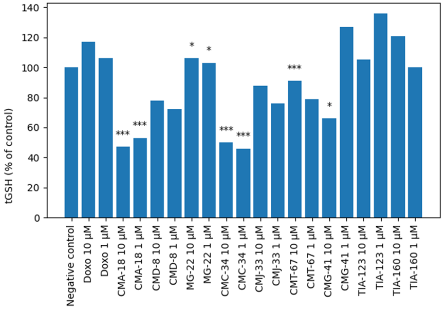
In the present study, DOXO (the positive control) at a concentration of 10.0 μmol/L induced a moderate increase in tGSH by 17% (Figure 1), GSH by 19% (Figure 2), and GSSG by 16% (Figure 3), with relative preservation of the GSH/GSSG ratio (+5%) (Figure 4), suggesting an adaptive antioxidant response. At 1.0 μmol/L, the effects were less pronounced, with a 10% decrease in the GSH/GSSG ratio (Figure 4), suggesting a mild dose-dependent redox imbalance relative to the negative control.
The benzothiazole derivatives CMA-18, CMD-8, and MG-22 produced distinct effects. CMA-18, at both tested concentrations, induced a marked reduction in tGSH by 47–53% and GSSG by 77–79%, together with a moderate decrease in GSH by 18–30%. Despite the depletion of total glutathione reserves, the GSH/GSSG ratio increased significantly by 212–312%, indicating predominance of the reduced form. However, the concurrent fall in tGSH suggests that this effect should be interpreted as redox modulation rather than unequivocal antioxidant protection.
CMD-8 produced moderate decreases in tGSH by 22–28% and GSSG by 42-52%, while maintaining GSH close to control values (2–3% variation), which led to an increase in the GSH/GSSG ratio by 71-95%. In contrast, MG-22 exhibited less pronounced effects, with tGSH values close to control (3-6% variation) and moderate changes in the GSH/GSSG ratio, namely − 17% at 1.0 μmol/L and +39% at 10.0 μmol/L, suggesting a limited impact on redox status. These findings indicate that benzothiazole Cu-thiosemicarbazones may limit glutathione oxidation, although their biological interpretation depends on the associated changes in total glutathione reserves.
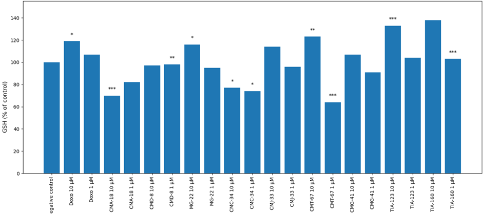
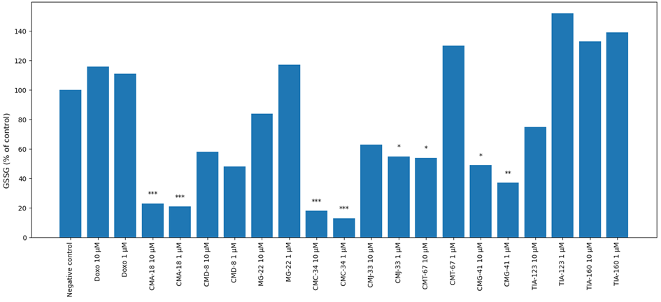
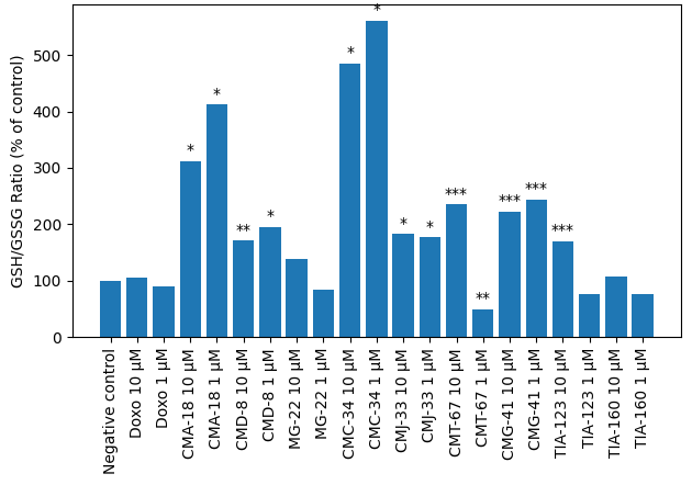
Phenyl-group Cu-thiosemicarbazones exhibited distinct effects at both concentrations. CMC-34 produced a significant reduction in tGSH by 45–50% and GSSG by 82–87%, accompanied by a moderate decrease in reduced GSH by 23–26%. This redistribution was associated with the greatest increases in the GSH/GSSG ratio (385–461%), suggesting a strong antioxidant effect and effective limitation of glutathione oxidation. CMJ-33 induced moderate decreases in tGSH by 12–24% and GSSG by 37–45%, with an increase in the ratio of 77–83%. CMT-67 showed a concentration-dependent effect: at 10.0 μmol/L, reduced GSH increased by 23% and the GSH/GSSG ratio by 135%, whereas at 1.0 μmol/L, GSH decreased by 36%, GSSG increased by 30%, and the ratio declined by 51%, suggesting a potential pro-oxidant effect at lower concentrations. Overall, phenyl Cu-thiosemicarbazones displayed the highest antioxidant potential, as evidenced by an increased GSH/GSSG ratio and reduced GSSG levels, indicating superior efficacy in maintaining a reductive intracellular environment, with the exception of CMT-67 at 1.0 μmol/L, which suggested a potential pro-oxidant effect (Figure 4).
Statistical evaluation of the allyl-group Cu-thiosemicarbazones showed that CMG-41 at 10.0 μmol/L reduced tGSH by 33% and GSSG by 51%, while maintaining reduced GSH at +7% and increasing the GSH/GSSG ratio by 122%. At 1.0 μmol/L, tGSH increased by 27%, whereas GSSG decreased by 63%, with the ratio reaching +144%, indicating substantial antioxidant efficiency. TIA-123, at both concentrations, induced increases in tGSH by 5–36% and GSH by 4–33%; however, at 1.0 μmol/L, an accumulation of GSSG (+52%) was observed, with GSH/GSSG ratio variations ranging from −23% to +70%, suggesting a biphasic effect on redox balance. Likewise, TIA-160 at 10.0 μmol/L increased tGSH by 21%, GSH by 38%, GSSG by 33%, and the GSH/GSSG ratio by 8%. At 1.0 μmol/L, tGSH remained at control levels, GSH increased by 3%, GSSG by 39%, while the GSH/GSSG ratio decreased by 23%. The allyl compounds (TIA-123 and TIA-160) thus demonstrated a dual effect, characterized by increased total glutathione reserves along with variable GSSG accumulation, indicating simultaneous activation of antioxidant mechanisms and oxidative processes. With the exception of CMG-41 at 10.0 µmol/L, the allyl compounds generally appeared to favor total glutathione accumulation and support an adaptive cellular response, although GSSG accumulation in some conditions indicates simultaneous activation of oxidative processes.
These findings suggest that structural modifications of substituents directly influence the ability to modulate redox balance, and that the structure–activity relationship plays a crucial role in determining the final biological effect.
In summary, the results demonstrate that the effects on redox status are dependent on both chemical structure and concentration. CMA-18, CMC-34, CMG-41, and CMT-67 at 10.0 μmol/L exhibited the most pronounced antioxidant potential, as evidenced by a significant increase in the GSH/GSSG ratio and a reduction in GSSG levels. These data support the hypothesis that structural modifications of benzothiazole, phenyl, and allyl Cu-thiosemicarbazones directly influence their ability to modulate cellular redox homeostasis.
Influence of Cu-thiosemicarbazones on glutathione metabolism and redox homeostasis
The present study shows that Cu-thiosemicarbazone compounds exert significant and distinct effects on glutathione metabolism, reflecting how the chemical structure of the compound may influence cellular redox homeostasis. Understanding these mechanisms is essential, since glutathione is the principal intracellular antioxidant and a major determinant of cellular vulnerability to OS. GSH acts by neutralizing ROS and maintaining the redox state of cysteine residues in proteins, whereas the GSH/GSSG ratio is a sensitive indicator of cellular OS. Low values are associated with ROS accumulation and cellular injury, whereas increased values suggest either a reduction in GSSG or an imbalance between the redox forms [22, 23].
DOXO (the positive control) induced moderate increases in tGSH and GSH without drastic changes in the GSH/GSSG ratio, confirming its role as a redox-modulating agent in this experimental system. In antitumor studies, DOXO has been shown to generate free radicals and increase OS through iron-dependent redox reactions; cells may respond by activating antioxidant defenses, including glutathione synthesis [24, 25].
The increases in tGSH, GSH, and GSSG observed in the DOXO-treated groups are consistent with data showing that this antineoplastic agent generates ROS through mitochondrial redox cycling and interactions with intracellular iron. Experimental evidence indicates that DOXO exposure may initially activate antioxidant defense mechanisms, followed by progressive depletion when oxidative pressure persists [24, 26]. The decrease in the GSH/GSSG ratio at the lower dose may reflect relative GSSG accumulation and impaired GSH regeneration, a phenomenon described in tumor-related oxidative imbalance [27].
The results obtained in this study highlight the differential modulation of cellular redox homeostasis by Cu-thiosemicarbazone compounds, reflected in changes in tGSH, GSH, GSSG, and the GSH/GSSG ratio. Compared with the negative control, the phenyl compounds (CMC-34, CMJ-33, CMT-67) produced the largest increases in the GSH/GSSG ratio (77-461%) and reduced GSSG levels, indicating limited glutathione oxidation. However, because CMC-34 and some other compounds also decreased tGSH, the observed ratio increase may partly reflect selective GSSG depletion or glutathione consumption rather than a purely cytoprotective effect.
The GSH/GSSG ratio as a marker of oxidative stress
In the scientific literature, the GSH/GSSG ratio is regarded as one of the most sensitive indicators of cellular redox status. A decrease in this ratio reflects increased OS and glutathione oxidation, whereas an increase indicates predominance of the reduced form and greater antioxidant capacity [28].
In the present study, several Cu-thiosemicarbazone compounds (for example CMA-18, CMC-34, CMG-41, and CMT-67) produced marked increases in the GSH/GSSG ratio (up to 461% relative to control). This behavior is comparable to observations reported for bioactive compounds capable of modulating glutathione metabolism and activating antioxidant mechanisms or inhibiting GSH oxidation [23]. Nevertheless, interpretation of this ratio requires simultaneous consideration of tGSH and GSSG, because a high ratio can occur even when total glutathione reserves are reduced.
Effects on tGSH and GSH levels
In the performed experiments, some compounds, including CMA-18 and CMC-34 produced significant decreases in tGSH and GSH. Similar phenomena have been reported in studies on agents that induce OS or interact directly with the glutathione system. Depletion of GSH is associated with increased cellular susceptibility to free radicals and disruption of redox homeostasis [29].
Other studies have likewise shown that low GSH levels or a decreased GSH/GSSG ratio are indicators of OS and occur frequently in tumor cells or under conditions of intense OS [30]. In this context, the reduction in tGSH observed for some derivatives may indicate either glutathione consumption in antioxidant reactions or conjugate formation with the tested compounds [11].
Compared with literature data, the changes observed in the present study indicate that Cu-thiosemicarbazone compounds may act through mechanisms related to disruption of glutathione metabolism and modulation of cellular OS. Numerous studies have shown that changes in GSH and GSSG levels represent sensitive indicators of the toxic or pharmacological effects of different compounds. According to Jones (2006), a decrease in the GSH/GSSG ratio indicates the onset of oxidative stress and accumulation of ROS, whereas an increase in the ratio reflects predominance of a reduced intracellular environment and enhanced antioxidant capacity [29].
Thus, the increase in the GSH/GSSG ratio observed for most compounds suggests maintenance of a more reduced redox state or stimulation of glutathione regeneration. Conversely, the decrease in tGSH observed for some structures may indicate glutathione consumption in detoxification reactions or direct interaction with the tested compounds. In this context, changes in tGSH, GSH, and GSSG levels observed across different studies are considered important indicators of the mechanisms of action of bioactive compounds. Some structures may induce oxidative stress through GSH consumption, whereas others may stimulate glutathione regeneration and increase the GSH/GSSG ratio, suggesting an antioxidant or cytoprotective effect.
The present study demonstrates that Cu-thiosemicarbazone coordination compounds differentially modulate glutathione-related redox parameters in a structure- and concentration-dependent manner. Several compounds produced marked changes in tGSH, GSSG, and the GSH/GSSG ratio, suggesting distinct interactions with cellular redox metabolism. However, because some compounds simultaneously reduced total glutathione reserves while increasing the GSH/GSSG ratio, these effects should be interpreted cautiously and cannot be considered unequivocally antioxidant. The comparative analysis identifies compounds with pronounced or moderate redox-modulating activity and supports the use of glutathione system parameters as informative markers for preliminary screening. Further studies are required to clarify the underlying mechanisms and to evaluate cytotoxic, pro-oxidant, or cytoprotective effects in relevant cellular models.
We express our gratitude to Academician Aurelian Gulea and his colleagues from the Advanced Materials Research Laboratory in Biopharmaceuticals at the State University of the Republic of Moldova for synthesizing the compounds.
Libretti S, Puckett Y. Physiology, homeostasis. In: StatPearls. Treasure Island (FL): StatPearls Publishing; 2023.
Le Gal K, Schmidt EE, Sayin VI. Cellular Redox Homeostasis. Antioxidants (Basel). 2021;10(9):1377. doi:10.3390/antiox10091377
Marengo B, Nitti M, Furfaro AL, et al. Redox Homeostasis and Cellular Antioxidant Systems: Crucial Players in Cancer Growth and Therapy. Oxid Med Cell Longev. 2016;2016:6235641. doi:10.1155/2016/6235641
Hileman EO, Liu J, Albitar M, Keating MJ, Huang P. Intrinsic oxidative stress in cancer cells: a biochemical basis for therapeutic selectivity. Cancer Chemother Pharmacol. 2004;53(3):209-219. doi:10.1007/s00280-003-0726-5
Behrend L, Henderson G, Zwacka RM. Reactive oxygen species in oncogenic transformation. Biochem Soc Trans. 2003;31(Pt 6):1441-1444. doi:10.1042/bst0311441
Waris G, Ahsan H. Reactive oxygen species: role in the development of cancer and various chronic conditions. J Carcinog. 2006;5:14. doi:10.1186/1477-3163-5-14
Chen TH, Wang HC, Chang CJ, Lee SY. Mitochondrial Glutathione in Cellular Redox Homeostasis and Disease Manifestation. Int J Mol Sci. 2024;25(2):1314. doi:10.3390/ijms25021314
Marrocco I, Altieri F, Peluso I. Measurement and Clinical Significance of Biomarkers of Oxidative Stress in Humans. Oxid Med Cell Longev. 2017;2017:6501046. doi:10.1155/2017/6501046
Garbuz O, Ceban E, Istrati D, Railean N, Toderas I, Gulea A. Thiosemicarbazone-Based Compounds: Cancer Cell Inhibitors with Antioxidant Properties. Molecules. 2025;30(9):2077. doi:10.3390/molecules30092077
Malarz K, Mrozek-Wilczkiewicz A, Serda M, Rejmund M, Polanski J, Musiol R. The role of oxidative stress in activity of anticancer thiosemicarbazones. Oncotarget. 2018;9(25):17689-17710. doi:10.18632/oncotarget.24844
Aquilano K, Baldelli S, Ciriolo MR. Glutathione: new roles in redox signaling for an old antioxidant. Front Pharmacol. 2014;5:196. doi:10.3389/fphar.2014.00196
Checa J, Aran JM. Reactive Oxygen Species: Drivers of Physiological and Pathological Processes. J Inflamm Res. 2020;13:1057-1073. doi:10.2147/JIR.S275595
Franco R, Cidlowski JA. Glutathione efflux and cell death. Antioxid Redox Signal. 2012;17(12):1694-1713. doi:10.1089/ars.2012.4553
Traverso N, Ricciarelli R, Nitti M, et al. Role of glutathione in cancer progression and chemoresistance. Oxid Med Cell Longev. 2013;2013:972913. doi:10.1155/2013/972913
Meyer AJ. The integration of glutathione homeostasis and redox signaling. J Plant Physiol. 2008;165(13):1390-1403. doi:10.1016/j.jplph.2007.10.015
Pantea V, Pavlovschi E, Stratulat S, Gulea A, Tagadiuc O, Gudumac V. Targeting Redox Balance: Antioxidant Effects of Thiosemicarbazones on Human Peripheral Blood. Mold J Health Sci. 2025;12(3):101-109. doi:10.52645/MJHS.2025.3.16
Puri KS, Suresh KR, Gogtay NJ, Thatte UM. Declaration of Helsinki, 2008: implications for stakeholders in research. J Postgrad Med. 2009;55(2):131-134. doi:10.4103/0022-3859.52846
Gulea A, Poirier D, Roy J, et al. In vitro antileukemia, antibacterial and antifungal activities of some 3d metal complexes: chemical synthesis and structure - activity relationships. J Enzyme Inhib Med Chem. 2008;23(6):806-818. doi:10.1080/14756360701743002
Pantea V, Pavlovschi E, Macari V, Tagadiuc O. Effects of copper(II) thiosemicarbazones on pro-oxidant and antioxidant system markers in erythrocytes. Farmacia. 2025;73(3):759-768. doi:10.31925/farmacia.2025.3.22
Ngamchuea K, Batchelor-McAuley C, Compton RG. Rapid Method for the Quantification of Reduced and Oxidized Glutathione in Human Plasma and Saliva. Anal Chem. 2017;89(5):2901-2908. doi:10.1021/acs.analchem.6b04186
Ryzhikov SI, Druzhinina IuG, Riabicheva TG, Varaksin NA. Standartizatsiia tekhniki opredeleniia produktsii tsitokinov kletkami krovi ex vivo [Standardization of technique for detection of blood cell cytokine production ex vivo]. Klin Lab Diagn. 2011;(11):49-53.
Forman HJ, Zhang H, Rinna A. Glutathione: overview of its protective roles, measurement, and biosynthesis. Mol Aspects Med. 2009;30(1-2):1-12. doi:10.1016/j.mam.2008.08.006
Townsend DM, Tew KD, Tapiero H. The importance of glutathione in human disease. Biomed Pharmacother. 2003;57(3-4):145-155. doi:10.1016/s0753-3322(03)00043-x
Minotti G, Menna P, Salvatorelli E, Cairo G, Gianni L. Anthracyclines: molecular advances and pharmacologic developments in antitumor activity and cardiotoxicity. Pharmacol Rev. 2004;56(2):185-229. doi:10.1124/pr.56.2.6
Octavia Y, Tocchetti CG, Gabrielson KL, Janssens S, Crijns HJ, Moens AL. Doxorubicin-induced cardiomyopathy: from molecular mechanisms to therapeutic strategies. J Mol Cell Cardiol. 2012;52(6):1213-1225. doi:10.1016/j.yjmcc.2012.03.006
Lee EJ, Jang WB, Choi J, et al. The Protective Role of Glutathione against Doxorubicin-Induced Cardiotoxicity in Human Cardiac Progenitor Cells. Int J Mol Sci. 2023;24(15):12070. doi:10.3390/ijms241512070
Zitka O, Skalickova S, Gumulec J, et al. Redox status expressed as GSH:GSSG ratio as a marker for oxidative stress in paediatric tumour patients. Oncol Lett. 2012;4(6):1247-1253. doi:10.3892/ol.2012.931
Jones DP. Redefining oxidative stress. Antioxid Redox Signal. 2006;8(9-10):1865-1879. doi:10.1089/ars.2006.8.1865
Ferreira-Cravo M, Moreira DC, Hermes-Lima M. Glutathione Depletion Disrupts Redox Homeostasis in an Anoxia-Tolerant Invertebrate. Antioxidants (Basel). 2023;12(6):1197. doi:10.3390/antiox12061197
Schoeneberger H, Belz K, Schenk B, Fulda S. Impairment of antioxidant defense via glutathione depletion sensitizes acute lymphoblastic leukemia cells for Smac mimetic-induced cell death. Oncogene. 2015;34(31):4032-4043. doi:10.1038/onc.2014.338
Citation: Pantea V, Pavlovschi E. In vitro modulation of cellular redox homeostasis by cu-thiosemicarbazone coordination compounds. Arta Medica. 2026;99(2):e2026007. doi: 10.5281/zenodo.21552732